Table of Contents >> Show >> Hide
- The back in one sentence
- Quick diagrams you can actually understand
- The bony framework: the spine is the “mast,” not the “sail”
- The muscle layers: the back has “movers” and “managers”
- Thoracolumbar fascia: the connective tissue “power belt”
- Nerves: why the back can cause symptoms in your arm or leg
- Movements and biomechanics: how the parts cooperate
- Common clinical tie-ins (the “why you care” section)
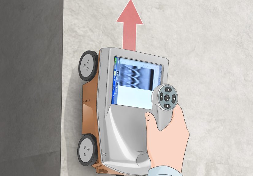
- How to read a back anatomy diagram like a pro
- Conclusion: the back is built for work, not for stillness
- Real-world experiences: what back anatomy feels like outside a textbook (≈)
Your back is basically the body’s “support team”: it holds you upright, lets you twist to grab the last slice of pizza,
and quietly absorbs the consequences of questionable chair choices. Anatomically, it’s a layered stack of bones, joints,
discs, ligaments, muscles, fascia, nerves, and blood vesselsorganized well enough to be elegant, but complex enough to
make anatomy students consider switching majors.
This guide breaks down back anatomy with simple diagrams (copy-friendly!), a practical overview of the spine and back
muscles, and the real-life “why it matters” connectionslike why a disc can cause leg symptoms, or why your shoulder
pain sometimes starts in your mid-back.
The back in one sentence
Back anatomy is the relationship between the vertebral column (bones and joints),
intervertebral discs (shock-absorbing pads), ligaments and fascia (connective “seatbelts” and “wrapping”),
and muscle layers (big movers plus small stabilizers), all wired together by spinal nerves and fed by segmental blood supply.
Quick diagrams you can actually understand
1) Posterior (back) view “map”
2) Cross-section “layer cake”
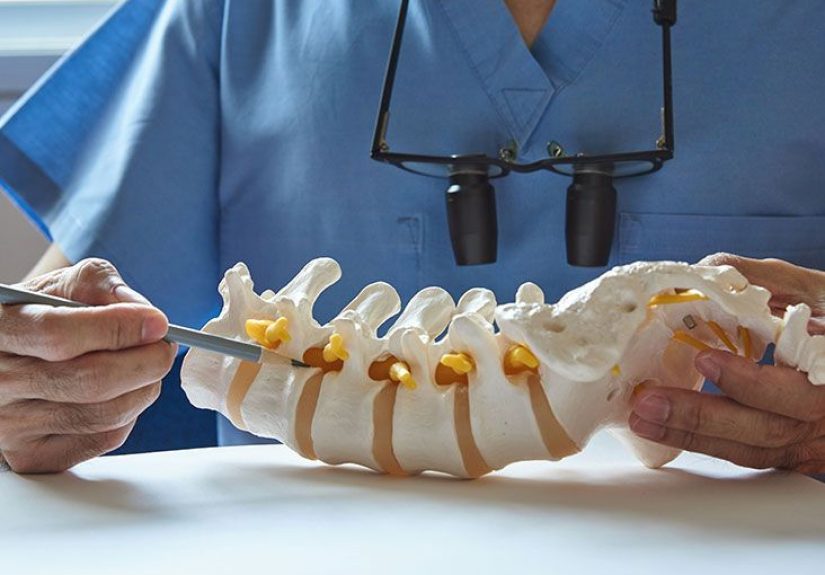
The bony framework: the spine is the “mast,” not the “sail”
The backbone of back anatomy (pun extremely intended) is the vertebral column. It’s built from
stacked vertebrae, separated by discs, connected by facet joints, and reinforced by ligaments. The structure protects
the spinal cord and nerve roots while still allowing motionan engineering compromise that works remarkably well,
until we ask it to tolerate eight hours of slumped texting.
Regions of the vertebral column
The spine is usually described in regions from top to bottom:
- Cervical spine (C1–C7): neck; mobility-focused.
- Thoracic spine (T1–T12): upper/mid-back; more stable thanks to rib attachments.
- Lumbar spine (L1–L5): lower back; major weight-bearing and motion.
- Sacrum (S1–S5 fused): forms the back wall of the pelvis.
- Coccyx (usually 3–5 fused segments): tailbone; small but not irrelevant (ask anyone who’s fallen on ice).
Intervertebral discs, joints, and “exit ramps” for nerves
Between most vertebrae sit intervertebral discs. Each disc has:
- Annulus fibrosus: a tough outer ring (think: durable tire).
- Nucleus pulposus: a gel-like center (think: shock-absorbing jelly donut filling).
In a disc herniation, some of that central material pushes outward and may irritate nearby nerve structures.
That’s how a spine problem can create symptoms far awaylike pain, numbness, or tingling down a leg.
Motion between vertebrae also involves facet joints (paired joints at the back of each vertebra).
These guide movement and share load. Nearby are the intervertebral foraminaopenings where spinal nerves exit.
If a disc bulges or surrounding tissues swell, the “exit ramp” can get crowded, and nerves tend to complain loudly.
The muscle layers: the back has “movers” and “managers”
A helpful way to understand back muscles is to separate them into:
(1) muscles that move the shoulder girdle/upper limb (more superficial),
and (2) muscles that stabilize and move the spine itself (deeper).
Superficial (extrinsic) back muscles: shoulder-and-arm influencers
These are the “headline” muscles people recognize in a back anatomy diagram because they’re big and close to the surface.
They mostly influence the shoulder girdle and upper limb position.
| Muscle | Where it sits | High-level job description |
|---|---|---|
| Trapezius | Upper back/neck region | Moves and stabilizes the scapula (elevation, retraction, rotation); posture support. |
| Latissimus dorsi | Broad lower back/side | Helps extend/adduct the arm; connects trunk to upper limb movement. |
| Rhomboids | Under trapezius | Retract and stabilize the scapula (think: “pin shoulder blades back”). |
| Levator scapulae | Neck to scapula | Elevates scapula; often involved in “tight neck/upper trap” feelings. |
Practical example: If your shoulders round forward at your desk, the scapulae drift, and these superficial muscles can become
overworked or underused in predictable ways. That’s why shoulder posture and mid-back comfort are basically roommates.
Intermediate back muscles: subtle helpers for breathing mechanics
The intermediate group (often described as part of the extrinsic category) includes muscles like the
serratus posterior superior and serratus posterior inferior. They attach to ribs and are often discussed as
assisting respiration mechanics or rib movementsmall players compared to the diaphragm, but relevant in thoracic region dynamics.
Intrinsic (deep) back muscles: posture, precision, and spine control
Intrinsic muscles are closer to the vertebral column and are the “managers” of spinal movementespecially posture, segment-by-segment stability,
and controlled rotation/extension. They’re not always glamorous, but they’re the reason you can stand still without collapsing like a folding chair.
- Splenius muscles (upper back/neck): contribute to head/neck extension and rotation; important in cervical mechanics.
-
Erector spinae group: the major “upright” muscle mass. Commonly described as three columns:
iliocostalis (more lateral), longissimus (intermediate), spinalis (more medial).
They extend the spine, assist lateral flexion, and help maintain normal spinal curves. -
Transversospinalis group: deeper stabilizers like multifidus and rotatores.
These are famous for segmental controlsmall movements, big importance (especially in low back stability). -
Minor segmental muscles: short muscles between vertebrae (like interspinales and intertransversarii)
that contribute to fine-tuning alignment.
Gym example: During a deadlift or even picking up a laundry basket, your erector spinae help resist spine flexion and control the return to upright.
Meanwhile, deeper muscles like multifidus contribute to segmental stabilitymore “precision steering,” less “big engine.”
Thoracolumbar fascia: the connective tissue “power belt”
The thoracolumbar fascia is a strong, multi-layered sheet of connective tissue in the lower and mid-back.
In a back anatomy diagram, it’s easy to ignore because it’s not a muscle or boneuntil you realize it helps transmit forces between the trunk and limbs,
provides broad attachment surfaces, and organizes layers of muscle.
Translation: it’s part of why the back can handle load transferlike when you brace to lift, push, or twist. If muscles are the “workers,” fascia is
the “infrastructure” that makes teamwork more efficient.
Nerves: why the back can cause symptoms in your arm or leg
Spinal cord, conus medullaris, and cauda equina
The spinal cord travels through the spinal canal and typically tapers around the upper lumbar region into the
conus medullaris. Below that, nerve roots continue downward as a bundle called the cauda equina
(“horse’s tail”anatomists were poets, apparently).
This arrangement matters clinically: compression or severe irritation of these structures can cause pain, weakness,
sensory changes, andmost urgentlybowel or bladder dysfunction.
Dorsal (posterior) rami: the wiring for intrinsic back muscles
Spinal nerves branch into rami. A key concept in back anatomy is that the dorsal/posterior rami primarily supply
the intrinsic (deep/true) muscles of the back and the skin overlying the back. That’s one reason deep back pain can feel
local and “stripe-like” on the skin.
Movements and biomechanics: how the parts cooperate
The spine moves in multiple directions, and different structures contribute depending on the motion and load:
- Flexion (bending forward): discs and ligaments resist excessive motion; muscles control the descent and return.
- Extension (bending backward): facet joints guide motion; erector spinae contribute strongly.
- Lateral flexion (side-bending): asymmetrical muscle activation and joint mechanics share the work.
- Rotation (twisting): thoracic region rotates more than lumbar; deep stabilizers help keep segments coordinated.
The big idea: your back isn’t just a column of bonesit’s a controlled, load-sharing system. Muscles provide force and stability,
discs and joints allow motion, ligaments and fascia provide passive support, and nerves coordinate it all.
Common clinical tie-ins (the “why you care” section)
Strain vs. disc irritation vs. stenosis
-
Muscle strain: often feels local, achy, and worse with specific movements or prolonged posture.
It can involve superficial or deep layerssometimes both, because your back loves a group project. -
Disc herniation/irritation: may cause radiating symptoms (like down a leg) if nerve roots are affected.
Sensations can include sharp pain, tingling, numbness, or weakness. -
Spinal stenosis: narrowing around neural structures can produce position-dependent symptoms (often worse with extension/standing,
sometimes improved with flexionyour mileage may vary and should be assessed clinically).
Red flags you don’t “walk off”
Because the cauda equina carries nerves for lower limb function and pelvic organs, a true cauda equina syndrome scenario can be urgent.
If someone develops new bowel/bladder dysfunction, saddle-area numbness, or rapidly progressive weakness, that’s not a “stretch it out” situation.
It’s a “get evaluated promptly” situation.
Posture: less about perfection, more about options
A common misconception is that there’s one “perfect” posture that saves your spine forever. A more realistic (and anatomy-friendly) idea:
the healthiest posture is usually the one you can change. Variety reduces repetitive stress, and movement keeps tissues from acting like they’ve
filed a formal complaint with HR.
How to read a back anatomy diagram like a pro
- Find landmarks first: spine midline, scapulae, rib cage outline, iliac crests (top of pelvis).
- Identify the layer: superficial diagrams show trapezius/latissimus; deeper diagrams show erector spinae/multifidus.
- Match muscle direction to function: fibers running upward/diagonal often relate to pulling or stabilizing in that direction.
- Look for “columns” near the spine: those are often intrinsic stabilizers rather than shoulder movers.
-
Connect nerves to symptoms: radiating pain suggests nerve involvement; local ache often suggests muscular or joint contribution.
(This is a clue, not a diagnosis.)
Conclusion: the back is built for work, not for stillness
A clear back anatomy overview comes down to layers and relationships:
bones and joints form the framework; discs and ligaments manage load and motion; muscles provide power and stability;
thoracolumbar fascia helps force transfer; and nerves make sure the whole system operates as one coordinated unit.
When you understand how a diagram maps onto those layers, you can interpret common problems more intelligently,
communicate better with clinicians or trainers, and make everyday choices that are kinder to your spinelike rotating tasks,
varying positions, and treating “core support” as a team sport, not a punishment.
Real-world experiences: what back anatomy feels like outside a textbook (≈)
One of the most common experiences people have when learning back anatomy is realizing they’ve been pointing to the “wrong” place for years.
Someone says “my lower back hurts,” and their hand lands on the back of the pelvis or even the upper glute. That’s not ignoranceit’s practical
human mapping. The good news is that anatomy gives you a better map: lumbar spine is above the pelvis, sacrum sits between the hip bones, and
muscles like the erector spinae and gluteals work together so closely that discomfort can blur the borders.
Another classic moment happens in the gym or during a move: you lift something “with your back,” feel a sudden tightness, and immediately bargain
with the universe. What’s often going on is that your back muscles are acting like the emergency brakes. They fire to prevent uncontrolled flexion,
to stabilize the spine, and to transmit force between your legs and your upper body. If your hips and core don’t share enough of the load, the
back tries to do everythinglike the coworker who “just handles it” until they finally snap and take a vacation.
People also discover that back anatomy is deeply connected to breathing and stress. When you’re anxious, your shoulders creep up, your neck feels
tight, and suddenly your upper trapezius is starring in a one-muscle Broadway show. Meanwhile, the thoracic spine may feel stiff because you’re not
moving your rib cage much. This is where the “layer” concept helps: superficial muscles can get cranky when deeper stabilizers aren’t doing their quiet
job, and posture becomes a symptom of the system rather than a single moral failing.
In clinics and physical therapy settings, a frequent experience is learning the difference between “pain” and “danger.” Many back issues are painful
but not structurally catastrophic. That doesn’t mean the pain is imaginaryjust that tissues can be sensitive without being broken. Understanding
discs, joints, and nerve pathways helps explain why symptoms can travel (like sciatica-like sensations) and why certain positions flare things up.
People often feel relieved when they realize a radiating symptom can be a nerve irritation pattern, not a mysterious curse.
And then there’s the “diagram-to-life” experience: you see an illustration of the cauda equina and suddenly understand why certain symptoms are treated
urgently. It’s sobering in a useful waylike reading the instruction manual before you break the machine. Anatomy doesn’t exist to scare you; it exists
to give you clear, actionable context. When people leave with one practical insight, it’s usually this: your back is designed to move and adapt, so
build habits that give it optionschange positions, strengthen gradually, and respect red flags when they show up.