Table of Contents >> Show >> Hide
- What “Endoscopy” Means in Ulcerative Colitis
- When Is Endoscopy Needed for UC?
- 1) To Diagnose UC (and Rule Out Look-Alikes)
- 2) To Map Disease Extent (How Far It Spreads)
- 3) To Measure Severity and Guide Treatment (“How Hot Is the Fire?”)
- 4) During a Suspected Flare or Worsening Symptoms
- 5) To Check Response to Treatment and Support Treat-to-Target Goals
- 6) Cancer Prevention: Dysplasia Surveillance in Long-Standing UC
- Types of Endoscopy Used in Ulcerative Colitis
- What Happens During a UC Endoscopy?
- What Results Might Show (And What They Mean)
- Risks, Complications, and When to Seek Urgent Help
- How to Make the Experience Easier (Without Becoming a Prep Influencer)
- Conclusion
- Real-Life Experiences: What UC Endoscopy Is Like (From the People Who’ve Been There)
If ulcerative colitis (UC) had a “behind-the-scenes” documentary, endoscopy would be the camera crew: quietly doing the hard work, occasionally making you question your life choices (hello, bowel prep), and ultimately revealing what’s really happening inside your colon. When symptoms flare or decisions get complicated, an endoscopic exam can turn guesses into evidence and help you and your GI team choose the next move with confidence.
This guide breaks down when UC endoscopy is needed, the main types of endoscopy used in UC, and exactly what happens during the procedurewith enough practical detail to feel prepared, but not so much that you start practicing deep breathing in the grocery aisle. (Save that for prep night.)
What “Endoscopy” Means in Ulcerative Colitis
In everyday UC care, “endoscopy” usually means a doctor uses a thin, flexible tube with a camera and light to look directly at the lining of your large intestine (the colon) and rectum. The big reason this matters: UC is defined by inflammation on the inside surface of the colon, and endoscopy is one of the most direct ways to see that inflammation, judge how severe it is, and take tissue samples (biopsies) when needed.
Think of symptoms as your body’s “push notifications.” Helpful, but not always specific. Endoscopy is the in-person meeting where you finally see the full thread.
When Is Endoscopy Needed for UC?
There’s no single schedule that fits everyone. Endoscopy is usually recommended when it will change what your care team does nextdiagnosis, treatment strategy, monitoring, or cancer prevention.
1) To Diagnose UC (and Rule Out Look-Alikes)
If you’re newly experiencing symptoms like persistent diarrhea, rectal bleeding, urgency, and abdominal cramping, doctors often use colonoscopy or flexible sigmoidoscopy with biopsies to confirm UC and exclude other causes. Stool tests and blood work may help rule out infections or other conditions, but biopsies taken during endoscopy can provide the defining evidence of UC-related inflammation.
Example: Two people can have the same symptomsbloody diarrhea and urgencybut one might have UC while the other has an infection or another form of colitis. Endoscopy plus biopsy helps clarify which “plot” you’re actually living in.
2) To Map Disease Extent (How Far It Spreads)
UC commonly starts in the rectum and may extend upward in a continuous pattern. Knowing how much of the colon is involved matters because it can influence medication choices, monitoring intensity, and long-term risk discussions.
- Proctitis: inflammation limited to the rectum
- Left-sided colitis: extends through the sigmoid/descending colon
- Extensive colitis (pancolitis): extends beyond the splenic flexure, possibly involving the entire colon
This “map” also helps your team interpret symptoms. For example, significant urgency with minimal stool volume might fit distal inflammation, while more frequent, larger-volume diarrhea may suggest broader involvement.
3) To Measure Severity and Guide Treatment (“How Hot Is the Fire?”)
Endoscopy doesn’t just say “inflammation: yes/no.” It can help grade severity using validated scoring systems such as the Mayo Endoscopic Score or the UC Endoscopic Index of Severity (UCEIS). In plain language, your doctor is looking for clues like: loss of normal blood vessel pattern, redness, friability (easy bleeding), erosions, and ulcers.
Severity matters because it influences decisions like whether topical therapy might be enough, whether oral medications should be stepped up, or whether hospitalization-level treatment is appropriate.
4) During a Suspected Flare or Worsening Symptoms
If symptoms return after a period of stability, your care team may recommend flexible sigmoidoscopy or colonoscopy to confirm whether inflammation is back, assess how intense it is, and rule out complications or infections that can mimic flares.
Importantly, not every symptom spike equals active inflammation. Some people experience ongoing symptoms from IBS-like overlap, infection, medication side effects, or residual sensitivity even when the lining has improved. Endoscopy can clarify what’s driving symptoms and prevent “treating the wrong problem.”
5) To Check Response to Treatment and Support Treat-to-Target Goals
Modern UC care often aims beyond “feeling better” and toward measurable inflammation controlsometimes called mucosal healing. An endoscopically healthier lining is associated with more durable remission and fewer complications over time.
In practice, many clinicians combine symptoms, biomarkers (like fecal calprotectin and CRP), and targeted endoscopy when decisions depend on objective proof of healing. Sometimes a shorter flexible sigmoidoscopy is enough for monitoring, especially if prior inflammation was mostly in the left colon.
6) Cancer Prevention: Dysplasia Surveillance in Long-Standing UC
People with long-standing colonic UC can have a higher risk of colorectal cancer than the general population, especially with extensive disease, ongoing inflammation, family history of colorectal cancer, or coexisting conditions like primary sclerosing cholangitis (PSC). Surveillance colonoscopy is designed to detect dysplasia (precancerous changes) early, when it’s most treatable.
Many expert recommendations suggest starting dysplasia screening colonoscopy about 8–10 years after diagnosis for colonic inflammatory bowel disease, and then repeating surveillance at intervals based on individual riskoften in the 1–5 year range depending on factors like inflammation history, PSC, prior dysplasia, and the quality of prior exams.
Types of Endoscopy Used in Ulcerative Colitis
Colonoscopy
What it examines: the rectum and the entire colon. Colonoscopy is commonly used for diagnosis (especially to assess full extent), to evaluate symptoms, and for dysplasia surveillance. During the exam, the colon is gently inflated with air or CO2 so the doctor can see the lining clearly, and biopsies can be taken.
Typical timing: often about 30–60 minutes for the procedure itself, plus recovery time afterward. Sedation or anesthesia is common, so most people remember very littleand are required to have someone else drive them home.
Flexible Sigmoidoscopy
What it examines: the rectum and lower colon (usually the sigmoid and descending colon). It’s often used when doctors need a quicker look at active inflammation, especially during a flare, and it can be useful in hospitalized patients.
Why it’s popular in UC care: it usually takes less time (often 10–20 minutes), may require less bowel prep than colonoscopy, and often doesn’t require sedation. It’s also sometimes preferred if the colon is severely inflamed and a full colonoscopy could be riskier.
Chromoendoscopy (Dye-Spray or “Virtual” Techniques)
When the goal is dysplasia surveillance, many specialists aim to optimize detection using high-definition scopes and enhanced imaging. Dye-spray chromoendoscopy uses a contrast dye applied to the colon lining to highlight subtle changes. Virtual chromoendoscopy (built-in digital contrast modes on modern scopes) can be used as an alternative in many settings with high-definition equipment.
The point isn’t to make your colon look artsyit’s to make tiny, flat lesions easier to spot so biopsies can be targeted and meaningful.
Biopsies: The “Receipts” of Endoscopy
Biopsies are small tissue samples taken painlessly through the scope. They help confirm diagnosis, assess microscopic inflammation, evaluate dysplasia, and distinguish UC from other conditions.
In surveillance exams, your doctor may take targeted biopsies from anything that looks suspicious. If enhanced imaging isn’t used, some protocols include additional “nontargeted” biopsies in previously inflamed areas to improve dysplasia detection.
What Happens During a UC Endoscopy?
While specific steps vary by facility, most UC endoscopy visits follow the same storyline: preparation, check-in, procedure, recovery, and results. The only unpredictable part is whether you will laugh at the nurse’s “no plans today, right?” joke. (Correct answer: “Only a nap.”)
Step 1: Before the Procedure (Scheduling and Safety Checks)
- Medication review: You’ll be asked about prescription meds, supplements, and especially blood thinners. The goal is to reduce bleeding risk while keeping you safe from clotting risks.
- Health history: Heart/lung conditions, sleep apnea, prior anesthesia reactions, pregnancy status, and allergies may affect sedation choices.
- Why you’re having the exam: Diagnosis, flare evaluation, monitoring response, or dysplasia surveillance can change the plan (where to biopsy, whether to use chromoendoscopy, etc.).
Step 2: Bowel Prep (The Part Everyone Talks About)
A clean colon is essential. Residual stool can hide inflammation or precancerous lesions, turning a valuable test into an expensive peekaboo game nobody asked for. Prep usually involves:
- Diet changes for a few days (often lower fiber), then a clear-liquid day before the exam
- Laxatives (liquid, pills, powder, and sometimes enemas), frequently split between the night before and the morning of the procedure
- Avoiding red or purple liquids because they can look like blood inside the colon
- Planning logistics: easy bathroom access, soft wipes, skin barrier cream, and a “this is my life now” playlist
Practical tip: If you struggle to finish prep due to nausea or side effects, contact the clinic. Incomplete prep is a common reason exams need to be repeated sooner than planned.
Step 3: Check-In and Sedation
For colonoscopy, most people receive IV sedation or anesthesia so they won’t feel pain and may not be awake for the procedure. Sedation can range from moderate sedation (“sleepy and relaxed”) to deep sedation, depending on patient factors and facility practice. Flexible sigmoidoscopy is often done without sedation, though options vary.
You’ll change into a gown, start an IV if needed, and meet the care team. Then you’ll be positioned on your side. Dignity takes a short coffee break, but professionalism stays in the room the whole time.
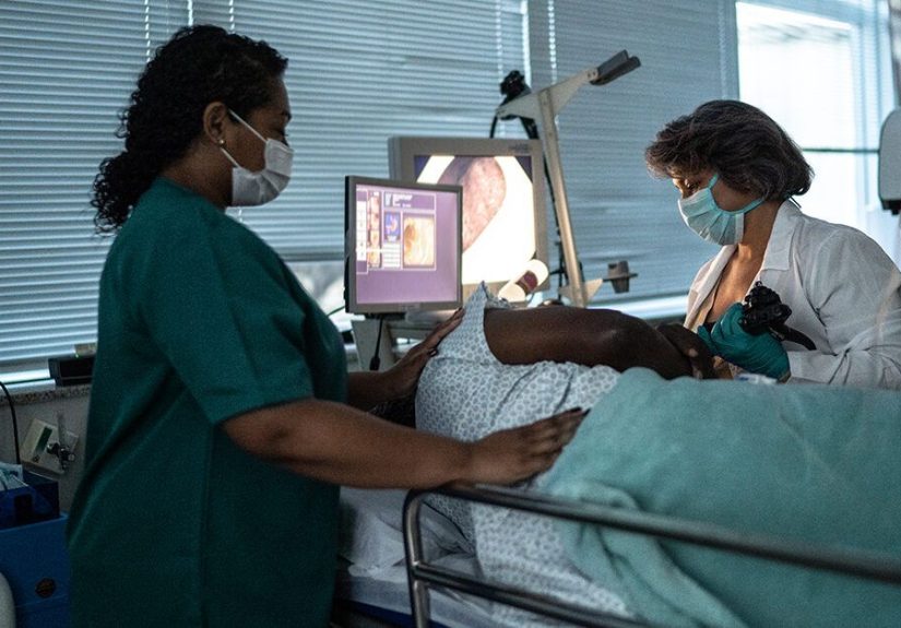
Step 4: The Procedure Itself
The scope is inserted through the anus and advanced through the rectum (and farther in colonoscopy). Air or CO2 gently inflates the colon for a clear view. The doctor examines the lining carefullyoften spending extra time on the way out because that’s where detail matters. If the purpose is surveillance, enhanced imaging and targeted biopsies may be used.
If the doctor sees inflamed areas, they may document:
- Location and extent (which segments are involved)
- Severity (validated scoring systems may be used)
- Features such as friability, erosions, ulcers, bleeding, or pseudopolyps
- Biopsies taken for diagnosis, inflammation grading, infection evaluation, or dysplasia assessment
Step 5: Recovery and Same-Day Findings
After colonoscopy, you’ll rest while sedation wears offoften 30–60 minutes at the facility, sometimes longer depending on sedation type. Mild cramping or bloating is common as the gas dissipates. If biopsies were taken, you might see a small amount of blood with the first bowel movement or two.
Many doctors can discuss what they saw right away. Biopsy results typically take several days or longer, depending on lab processing.
What Results Might Show (And What They Mean)
Active Inflammation vs. Healing
Endoscopic findings can range from a normal-looking lining to severe ulceration. Healing doesn’t always mean “perfect,” but improvement often supports staying the course. On the other hand, persistent ulcers or widespread friability may prompt medication changes even if symptoms are only “moderately annoying.”
Why Biopsy Results Matter Even When the Camera Looks Okay
Sometimes the colon lining looks much better, but microscopic inflammation remains. That can inform risk discussions and monitoring strategy, especially if you’ve had frequent flares or prior dysplasia. Your doctor may talk about clinical remission (symptoms), endoscopic remission (what they see), and sometimes histologic activity (microscope-level inflammation).
Dysplasia Findings
Dysplasia is not cancer, but it can be a warning sign. If dysplasia is found, next steps depend on whether it’s visible and removable, how confident the endoscopist is about complete resection, and whether there are additional concerning findings. In many cases, carefully performed repeat exams with advanced visualization are used to confirm findings and guide management.
Risks, Complications, and When to Seek Urgent Help
Endoscopy is generally safe, but no medical procedure is risk-free. Risks can be higher in certain situations, such as severe colitis or when therapeutic interventions are performed. Commonly discussed risks include:
- Bleeding (more likely after polyp removal; mild bleeding after biopsy can occur)
- Perforation (a tear in the colon wall; rare but serious)
- Sedation-related issues (breathing or heart complications, especially in higher-risk patients)
- Post-procedure discomfort (bloating, cramping, temporary changes in bowel habits)
Seek urgent care after an endoscopy if you develop severe or worsening abdominal pain, fever, repeated vomiting, heavy or persistent bleeding, dizziness, fainting, or significant weakness. Clinics typically provide a clear “call us vs. go to the ER” instruction sheetfollow it.
How to Make the Experience Easier (Without Becoming a Prep Influencer)
Prep Tips That Actually Help
- Chill the prep (cold tastes better), and use a straw to bypass some taste buds.
- Split-dose if prescribed: many regimens are designed to improve cleanliness and visibility.
- Protect your skin: barrier cream + soft wipes can be a game-changer.
- Clear liquids with electrolytes can help you feel less wiped out.
- Set up your “bathroom base camp”: phone charger, reading material, and a sense of humor.
Questions Worth Asking Your GI Team
- Is this exam for diagnosis, flare assessment, monitoring, or dysplasia surveillance?
- Should this be a colonoscopy or flexible sigmoidoscopy?
- Will you use chromoendoscopy or high-definition imaging for surveillance?
- Will biopsies be taken even if everything looks normal?
- How will results affect my treatment plan?
- When should I expect biopsy results, and how will I receive them?
Conclusion
UC endoscopy can feel intimidating, but it’s one of the most useful tools in ulcerative colitis careespecially when the goal is accurate diagnosis, smart treatment decisions, and long-term cancer prevention. Whether you’re having a quick flexible sigmoidoscopy during a flare or a detailed surveillance colonoscopy with enhanced imaging, the aim is the same: get clear information, reduce uncertainty, and help you stay in remission longer.
And yes, bowel prep is a nuisance. But it’s also the reason the exam can do what it’s supposed to dospot inflammation, confirm healing, and detect subtle changes early. In UC care, that clarity is worth a temporarily empty social calendar.
Real-Life Experiences: What UC Endoscopy Is Like (From the People Who’ve Been There)
Clinical explanations are helpful, but let’s be honest: the emotional experience of endoscopy deserves its own spotlight. Many people with UC describe the process as a weird mix of “no big deal” and “why is my life a bathroom-based mini-series?” Here are common, relatable themes patients shareespecially those who’ve done this more than once.
The “Prep Night” Reality Check
Most people don’t fear the scope as much as they fear the prep. The prep is disruptive, repetitive, and not exactly glamorous. Many say the first surprise is how long it takes. It’s not one dramatic moment; it’s an evening of frequent trips and the occasional, “Is my body producing liquids I didn’t know existed?”
People who’ve had multiple colonoscopies often develop a personal prep strategy: they keep the prep cold, pick a clear beverage they don’t love (so they won’t hate it later), and stock up on skin-protecting supplies. The most consistent “I wish someone told me” tip is about preventing irritation earlywaiting until your skin is already angry is like waiting to buy an umbrella until you’re soaked.
Showtime: Anxiety Before the Procedure
Even when you logically know endoscopy is routine, it can still trigger anxietyespecially if your last flare was rough or if you’re worried about dysplasia surveillance. Patients often say the worst part is the lead-up: you’re hungry, a bit dehydrated, and mentally rehearsing every possible outcome. Many find it calming to name the purpose of the test in one sentence: “We’re checking inflammation,” or “This is surveillance to keep me safe long-term.” That simple framing can reduce the sense of doom.
The Procedure Itself: “Wait… We’re Done?”
For sedated colonoscopy, a common experience is waking up and feeling like time teleported. People joke that it’s the best nap they didn’t planfollowed immediately by the world’s most enthusiastic offer of crackers and juice. Some feel bloated or crampy for a short time afterward because of the air used during the exam, but many are surprised that it’s milder than expected.
For flexible sigmoidoscopy without sedation, patients often describe it as uncomfortable but quick. The discomfort can feel like pressure or cramping, and some compare it to intense gas pains that pass once the procedure ends. The tradeoff is that you usually recover faster and can return to normal activities sooner.
Waiting for Biopsy Results: The Mental Marathon
Another shared experience: the waiting period for pathology results can feel longer than it actually is. Even if the doctor says, “Things look better,” people sometimes worry about what the biopsies might reveal. Many cope by planning something gentle for the next couple of dayswork, a favorite show, a low-stakes outinganything that prevents spiraling while they wait. It’s also common to write down questions right away, because post-sedation memory can be fuzzy.
What People Wish They’d Asked
Looking back, many UC patients say they wished they’d asked more specific questions: “What part of my colon was inflamed?” “How severe was it?” “Did you use a scoring system?” “Do these findings change my surveillance interval?” “Do we have a target for healing?” Knowing the answers can help people feel more in control and better prepared for the next steps.
The Unexpected Upside: Relief Through Certainty
Perhaps the most important “experience” theme is relief. Even when results show active disease, people often feel calmer once uncertainty is replaced with a plan. A scope can confirm healing when symptoms are confusing, or it can validate that symptoms have a real inflammatory driver. Either way, many patients describe endoscopy as a hard day that leads to clearer decisionsespecially when UC is unpredictable.
If you’re heading into an endoscopy soon, you’re not alone in feeling annoyed, nervous, or both. But most people come out the other side thinking some version of: “That wasn’t fun… but it was manageable, and it gave us answers.” In UC, answers are powerand power is how you get more good days on the calendar.