Table of Contents >> Show >> Hide
- What “AI Diagnosing Cancer” Actually Means
- Where AI Is Helping Diagnose Cancer Today
- 1) Breast cancer screening: smarter reads, saner workloads
- 2) Pathology: a second set of (digital) eyes on biopsy slides
- 3) Colonoscopy: real-time detection of polyps that can become cancer
- 4) Lung nodules and radiology triage: finding the “needle” earlier
- 5) Risk prediction: not diagnosis, but earlier warnings
- FDA, Regulation, and Why “Cleared” Isn’t the Same as “Perfect”
- What Makes AI Good at Diagnosing Cancer (and What Makes It Fail)
- How Hospitals Should Implement AI for Cancer Diagnosis
- What Patients Can Ask (Without Needing a Data Science Degree)
- The Bottom Line
- Real-World Experiences Related to AI Diagnosing Cancer (Extra Section)
- Experience #1: The radiologist who gains timeand loses patience for noise
- Experience #2: The pathologist who treats AI like a spell-checker (with humility)
- Experience #3: The patient who hears “AI helped” and wonders what that means for them
- Experience #4: The operations team that learns AI is a living system
- Experience #5: The “trust gap” phaseand how teams close it
- Conclusion
If you’ve ever stared at a blurry photo and said, “Enhance!” like you’re starring in a crime show,
you already understand the vibe of AI in cancer diagnosisexcept in real hospitals, the goal isn’t drama.
It’s earlier detection, fewer misses, fewer false alarms, and a workflow that doesn’t melt down when the scan volume spikes.
“AI diagnosing cancer” doesn’t mean a robot strolling into the exam room with a clipboard and an attitude.
In practice, AI is usually software that helps clinicians interpret complex datamedical images (mammograms, CTs, MRIs),
digital pathology slides, endoscopy video, and sometimes combinations of images + clinical data.
Think of it less as a replacement for a specialist and more like a tireless assistant that never needs coffee
and doesn’t get distracted halfway through the 400th image of the day.
This article breaks down where AI is already showing real value, where the hype outpaces the evidence,
and what “good” looks like when hospitals deploy these tools responsiblywithout turning cancer diagnosis into a guessing game.
What “AI Diagnosing Cancer” Actually Means
Most clinical AI is decision support, not a final verdict
In cancer care, AI commonly shows up in two places:
- Detection: “There’s something here worth a closer look.”
- Characterization: “This pattern looks more like X than Y,” often paired with a confidence score or heatmap.
The clinician remains responsible for interpretation. Many AI tools are built to flag suspicious regions,
triage cases, or improve consistencyespecially when humans are under time pressure.
AI learns patterns from examples (and the examples matter)
Modern medical AI is typically trained on large sets of labeled data: images or slides paired with “ground truth”
(biopsy results, expert consensus reads, follow-up outcomes). If the training data is skewedsay, mostly one device type,
one patient population, or one clinical settingperformance can drop in the real world.
So the story of AI diagnosing cancer is, in part, the story of data quality, diversity, and validation discipline.
Where AI Is Helping Diagnose Cancer Today
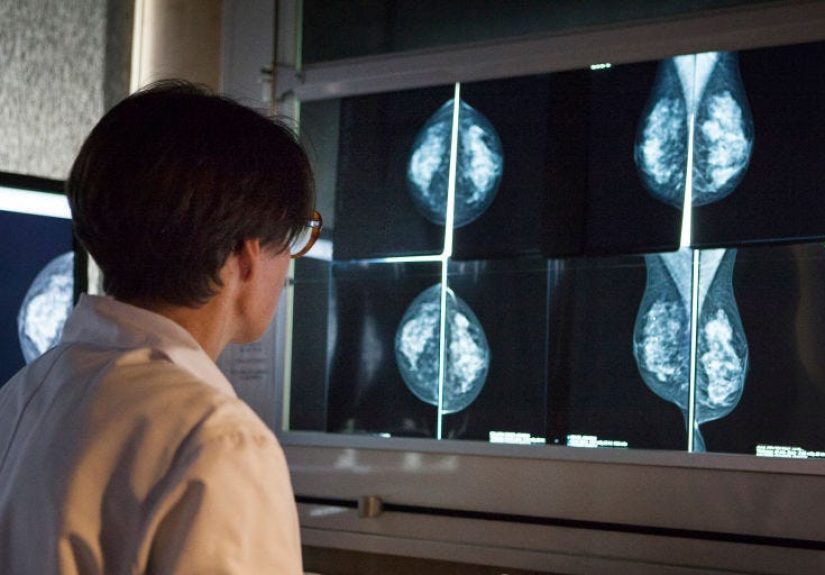
1) Breast cancer screening: smarter reads, saner workloads
Mammography is one of the most active areas for AI. Screening programs create huge image volumes, and subtle findings can be missed.
AI systems can highlight suspicious regions, score risk, and help radiologists prioritize tough cases.
In some large screening settings, AI has been studied as a way to maintain or improve detection while reducing workload.
The practical win here isn’t “AI beats doctors.” It’s “AI + doctors can be better than either alone,”
especially when the workflow is designed so the human-AI team plays to each other’s strengths.
2) Pathology: a second set of (digital) eyes on biopsy slides
Pathologists often diagnose cancer by examining tissue under a microscope.
As labs move toward whole-slide imaging (high-resolution digital scans of pathology slides),
AI can help find tiny foci of cancer that are easy to overlook, especially in high-volume settings.
A concrete example: FDA-authorized AI tools in pathology have been cleared to assist with detecting suspicious areas
in prostate biopsy slidessupporting pathologists as a quality-control backstop rather than replacing their expertise.
The tool can highlight regions that deserve a second look, which is particularly useful when the malignant area is small
and visually subtle.
3) Colonoscopy: real-time detection of polyps that can become cancer
Colorectal cancer often develops from precancerous polyps. During colonoscopy, AI-powered computer-aided detection (CADe)
can flag possible polyps in real time, nudging the clinician when the video feed shows something suspicious.
Multiple clinical studies have reported that AI-assisted colonoscopy can increase polyp and adenoma detection versus standard practice,
often with only a small increase in procedure time. That matters because higher adenoma detection is associated with lower future colorectal cancer risk.
4) Lung nodules and radiology triage: finding the “needle” earlier
CT imaging can detect lung nodules, but nodules vary widely, and clinical context matters.
AI tools can assist by marking nodules, measuring growth, and helping standardize follow-up recommendations.
There’s also growing interest in using AI platforms to expand early detection capacity to underserved settings,
where specialist availability may be limited.
5) Risk prediction: not diagnosis, but earlier warnings
Some AI models analyze “normal” or near-normal imaging to estimate future cancer risk.
For example, subtle texture patterns in mammograms may correlate with long-term breast cancer risk.
This isn’t a diagnosisbut it can guide screening intensity (who might benefit from supplemental imaging, shorter intervals, or closer monitoring).
The promise is targeted prevention: using smarter risk stratification to catch cancer earlieror keep it from becoming advanced.
FDA, Regulation, and Why “Cleared” Isn’t the Same as “Perfect”
Software as a Medical Device (SaMD) has rulesbecause it should
In the U.S., many clinical AI tools fall under “Software as a Medical Device.”
The FDA has built an evolving framework for reviewing AI/ML-based SaMD,
including action plans and guidance that emphasize a total product lifecycle approachwhat happens before deployment
(validation, intended use, safety) and after deployment (monitoring, updates, performance over time).
“FDA-cleared” means evaluated for a specific use in a specific way
A critical detail: an AI tool is evaluated for an intended use (for example, “as a concurrent reading aid” for screening mammography).
That means success depends on whether the tool is used as intendedon compatible equipment, in the right clinical workflow,
by trained professionals who understand what the output means and what it doesn’t.
What Makes AI Good at Diagnosing Cancer (and What Makes It Fail)
The upside: speed, consistency, and fewer misses
- Consistency: AI can reduce variation in readsespecially in repetitive tasks where fatigue is real.
- Prioritization: Triage models can push high-risk cases to the front of the line.
- Decision support: Heatmaps and scores can focus attention on areas that deserve a careful second look.
- Access: In low-resource or rural settings, AI may help extend specialist-level assistance where it’s scarce.
The hard parts: bias, drift, and “automation complacency”
The failure modes are just as important as the success stories:
-
Bias and uneven performance: A model trained on limited populations can underperform for others.
In cancer diagnosis, that’s not just a technical issueit’s an equity issue. -
Distribution shift and drift: Change the scanner, the staining method, the patient mix, or the protocol,
and performance can slide unless you monitor and recalibrate. -
False positives: Over-alerting wastes time and can lead to unnecessary follow-up.
(Also, nothing ruins a clinician’s day like an algorithm that cries wolf every 90 seconds.) -
False negatives: Missing a cancer is the nightmare scenario. Tools must be evaluated not only for average accuracy
but for clinically meaningful “misses,” especially aggressive cancers. -
Overreliance (“deskilling”): If clinicians lean too hard on AI, they may lose sharpness when the tool isn’t available.
That’s not an argument against AIit’s an argument for smart training and safeguards.
How Hospitals Should Implement AI for Cancer Diagnosis
1) Governance first: decide what “good” means locally
Responsible deployment starts with governance: selecting the right tool for the right job,
defining accountability, and documenting intended use. In imaging departments, professional guidance emphasizes
infrastructure, local acceptance testing, performance monitoring, and privacy considerations.
2) Local validation: “Works in a paper” is not “works here”
Before using AI in patient care, health systems should validate performance on local data:
the scanners they use, the patient demographics they serve, and the workflows clinicians actually follow.
If the model output changes behavior (for better or worse), that’s part of the evaluationnot an afterthought.
3) Monitor continuously: treat AI like a clinical instrument
AI isn’t a one-and-done purchase. Hospitals need post-deployment monitoring:
utilization rates, error patterns, data drift, clinician feedback, and periodic re-audits.
If performance degrades, you need a planupdate, recalibrate, restrict use, or pull it.
4) Train the humans: the best model still needs a competent team
Clinicians must understand:
- What the AI is designed to do (and what it is not designed to do)
- How to interpret outputs (scores, heatmaps, prompts)
- Common failure cases
- When to override the AIand how to document it
The goal is not blind trust. The goal is calibrated trust: using AI where it helps, questioning it where it’s weak,
and always keeping clinical judgment in the driver’s seat.
What Patients Can Ask (Without Needing a Data Science Degree)
If you’re a patient and you hear “we use AI,” you’re allowed to ask follow-ups. Helpful questions include:
- Is the AI tool FDA-cleared for this use? (And what exactly is that use?)
- Does a specialist still review the results? (They should.)
- How does the tool affect false alarms or missed findings?
- How is my data protected? (Privacy and security policies should be clear.)
- What happens if the AI and the clinician disagree? (There should be a process.)
AI can be a meaningful upgrade to carebut it should come with transparency, oversight, and measurable benefit,
not just buzzwords.
The Bottom Line
AI diagnosing cancer is realbut it’s not magic, and it’s not a sci-fi replacement for clinicians.
The strongest use cases are the ones you’d expect: image-heavy, pattern-driven workflows where fatigue, volume,
and subtle findings create opportunities for misses. Breast imaging, pathology slide review, and colonoscopy support
are leading examples.
The next chapter isn’t just “build better models.” It’s “build better systems”:
governance, validation, monitoring, training, and equitable performance across populations.
When that happens, AI becomes what it should have been all along: a practical tool that helps humans diagnose cancer earlier,
more consistently, and with fewer “how did we miss that?” moments.
Real-World Experiences Related to AI Diagnosing Cancer (Extra Section)
The most interesting stories about AI in cancer diagnosis aren’t the flashy headlines. They’re the small,
day-to-day moments in clinics where the tool either quietly helpsor quietly causes chaos.
Here are common, real-world experience patterns clinicians and patients describe when AI enters the workflow.
(These are not personal medical stories or guaranteesthink of them as “what it feels like in practice.”)
Experience #1: The radiologist who gains timeand loses patience for noise
In breast imaging, radiologists often describe the best AI as the kind they almost forget is there.
The model highlights a subtle region that genuinely deserves attention, or it helps downgrade an obviously normal study,
reducing cognitive load. Over time, the radiologist notices fewer “needle-in-a-haystack” misses and a smoother work rhythm.
But the same radiologists will also tell you: if the tool is too chatty, they’ll stop listening.
When AI flags too many harmless findings, it becomes background static.
Some departments tweak thresholds, refine protocols, and retrain staff so the AI alerts align with how clinicians actually work.
The lesson: adoption is less about algorithm brilliance and more about signal-to-noise management.
Experience #2: The pathologist who treats AI like a spell-checker (with humility)
In digital pathology, AI often feels like spell-check for cancer detection: it doesn’t write the essay,
but it circles the parts you might want to reread.
Pathologists describe using AI as a “final sweep” on prostate biopsy cases or other slide-heavy work:
first they review normally, then they glance at the AI heatmap to confirm nothing tiny is hiding in the corner.
What surprises some new users is how much trust depends on transparency.
If the tool highlights regions that “make sense” visually, pathologists build calibrated confidence.
If it highlights weird artifactsfolds in tissue, staining oddities, out-of-focus areasconfidence drops fast.
Successful labs keep feedback loops tight: pathologists report failure cases, the team tracks patterns,
and leadership decides whether the tool needs adjustment, better scanning standards, or narrower use.
Experience #3: The patient who hears “AI helped” and wonders what that means for them
Patients’ experiences tend to be emotionally straightforward: cancer testing is stressful,
and “AI” can sound either comforting (“extra help!”) or unsettling (“a computer decided?”).
Clinicians who explain AI well usually say something like:
“This tool helps us spot subtle patterns, but your results were reviewed by specialists.
We use it to reduce misses and improve consistency.”
Patients also report that transparency matters more than technical detail.
They don’t need a lecture on neural networks; they want reassurance about oversight, accuracy, and next steps.
The best conversations focus on outcomes: whether the tool improved detection, reduced delays,
or guided a follow-up plan with clear reasoning.
Experience #4: The operations team that learns AI is a living system
Hospital IT and clinical operations teams quickly discover that AI is not “install and forget.”
A scanner upgrade, new imaging protocol, or workflow change can shift the data the model sees.
Without monitoring, performance can driftsometimes subtly.
Teams that succeed treat AI like a clinical instrument that needs quality checks:
dashboards for usage, periodic audits, and clear escalation paths when performance looks off.
They also learn a very practical truth: if the AI output isn’t integrated cleanly into existing tools
(PACS viewers, pathology workstations, endoscopy platforms), clinicians won’t use it consistently,
and inconsistent use makes it harder to measure real benefit.
Experience #5: The “trust gap” phaseand how teams close it
Many sites go through a trust gap early on. Radiologists or pathologists may say, “Show me the evidenceon our patients.”
That skepticism can be healthy. The fastest way to close the gap is a structured pilot:
define metrics, compare performance with and without AI support, capture edge cases,
and be honest about trade-offs (like slightly more flagged findings in exchange for fewer misses).
When teams handle implementation with humilitytesting locally, monitoring continuously, and training thoughtfully
AI becomes less of a buzzword and more of a dependable teammate. Not a miracle worker. Not a menace.
Just a tool that, when used properly, can help clinicians diagnose cancer earlier and more consistently.
Conclusion
AI diagnosing cancer is already changing how clinicians read images, review slides, and detect precancerous findings.
The biggest wins come from well-defined roles: AI as assistive decision support, humans as accountable experts.
If you remember one thing, make it this:
the “best” AI isn’t the one with the flashiest demoit’s the one that improves real clinical outcomes,
stays reliable over time, and earns trust through transparency and monitoring.