Table of Contents >> Show >> Hide
- Quick Navigation
- What “Soft Tissue Sarcoma of the Foot” Actually Means
- Why the Foot Is a Tricky Neighborhood for Tumors
- Common Soft Tissue Sarcoma Types Seen in the Foot and Ankle
- Symptoms: When a “Foot Bump” Isn’t Just a Foot Bump
- Diagnosis: Imaging, Biopsy, and Why the Order Matters
- Staging & Grading: The Numbers That Drive Decisions
- Treatment Options for Soft Tissue Sarcoma of the Foot
- Recovery, Rehab, and Follow-Up: What Happens After Treatment
- Questions to Ask Your Sarcoma Team
- When to Seek Evaluation Quickly
- Conclusion
- Real-World Experiences: What the Journey Can Feel Like (Extra )
- SEO Tags (JSON)
Finding a weird lump on your foot can feel like discovering a surprise topping on your pizza: sometimes it’s harmless,
sometimes it’s… not the vibe. Most foot bumps are benign (ganglion cysts, plantar fibromas, lipomas, inflamed bursae),
but a small number are cancers called soft tissue sarcomas. Because the foot is basically a tightly packed
suitcase of tendons, nerves, blood vessels, and tiny bones, even a “small” tumor can cause outsized troubleand treatment
needs to be planned with extra care.
This guide breaks down what soft tissue sarcoma of the foot is, what symptoms to watch for, how diagnosis works
(including why the biopsy approach matters a lot), and what treatment and recovery can look like. It’s informationalnot a substitute
for medical carebut it will help you ask smarter questions and spot red flags earlier.
What “Soft Tissue Sarcoma of the Foot” Actually Means
Soft tissue sarcoma is a broad category of rare cancers that start in the body’s connective tissuesthings like
muscle, fat, fibrous tissue, blood vessels, and the linings around tendons and joints. “Of the foot” simply means the tumor is located
in the foot (or sometimes the ankle) rather than more common locations like the thigh or upper arm.
Two important realities can both be true at once:
- Most foot lumps are not cancer. Benign masses are far more common.
- When it is sarcoma, early expert evaluation matters. Delays and “let’s just remove it quickly” can make treatment harder.
Sarcomas are uncommon, and there are many subtypes. That’s why you’ll often hear doctors emphasize care at a
sarcoma center or by a multidisciplinary team (surgeons, oncologists, radiation specialists, pathologists,
radiologists, and often plastic/reconstructive experts).
Why the Foot Is a Tricky Neighborhood for Tumors
The foot has very little “extra space.” A mass that might quietly expand in a thigh has fewer places to hide in the footyet it can still be
missed because it’s easy to blame pain or swelling on shoes, plantar fasciitis, sports injuries, or that one pair of boots you refuse to retire.
What makes foot sarcomas uniquely challenging?
- Tight compartments: Tumors can wrap around tendons, nerves, or vessels sooner.
- Skin and soft tissue coverage is limited: After tumor removal, closing the wound may require flaps or grafts.
- Function is everything: Even small changes in the foot can affect gait, balance, and long-term mobility.
- Margins can be hard: Surgeons aim for a “wide” removal with a rim of healthy tissue, but anatomy can limit how wide is possible.
None of this means good outcomes aren’t achievablemany people do very well. It just means planning is not the place for improvisation.
In sarcoma care, the first procedure is often the most important one to get right.
Common Soft Tissue Sarcoma Types Seen in the Foot and Ankle
Sarcoma subtypes vary by age, location, and tissue origin. In the foot and ankle, clinicians commonly think about a few specific players:
Synovial sarcoma
Despite the name, it doesn’t have to arise from “synovium.” It often appears near joints and tendon sheaths, including the foot and ankle.
It may grow slowly and be mistaken for a benign lump early on.
Clear cell sarcoma
A rare tumor historically associated with tendons and aponeuroses (connective tissue structures), and it has a reputation for showing up in the
foot/ankle region. Because it can resemble other conditions under the microscope, expert pathology review is valuable.
Epithelioid sarcoma (distal type)
This subtype can appear in the hands and feet. It may look deceptively like a benign skin or soft tissue problem at first.
Other possibilities
Depending on imaging and biopsy results, doctors may also consider tumors such as malignant peripheral nerve sheath tumor, leiomyosarcoma,
liposarcoma (less common in the foot), undifferentiated pleomorphic sarcoma, angiosarcoma, and others.
The takeaway: “sarcoma” is not one disease. Subtype influences treatment decisions, recurrence risk, and follow-up strategy.
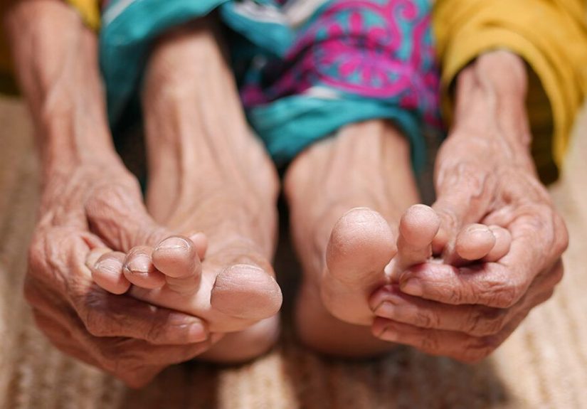
Symptoms: When a “Foot Bump” Isn’t Just a Foot Bump
The most common early sign of soft tissue sarcoma is a lump or swelling. In the foot, it might show up on the arch, the sole,
the top of the foot, between metatarsals, or near the anklesometimes painless, sometimes tender, sometimes only noticeable because your shoe suddenly
feels like it’s negotiating with you.
Red flags that deserve medical evaluation
- Growing over weeks or months (growth is one of the biggest warning signs)
- Larger size (commonly cited concern is around 5 cm / 2 inches, though smaller can still be serious in the foot)
- Deep location (feels “attached,” not freely movable under the skin)
- Persistent pain, tingling, or numbness (pressure on nerves)
- Recurrence after removal (“it came back” should never be ignored)
- An “injury” lump that doesn’t resolve (a presumed hematoma that sticks around deserves a closer look)
Common benign look-alikes (aka: why this gets missed)
Many non-cancerous conditions can mimic a sarcoma:
ganglion cysts, plantar fibromatosis, Morton’s neuroma, lipomas, tendon sheath tumors, bursitis, or scar tissue.
That’s why clinicians rely on imaging + biopsy rather than “vibes” to make the call.
Diagnosis: Imaging, Biopsy, and Why the Order Matters
Diagnosis usually follows a structured path: history and exam → imaging → biopsy → staging work-up. The goal is to identify the tumor accurately
and plan treatment without accidentally making later surgery harder.
Step 1: Imaging (MRI is often the star)
For foot and ankle soft tissue tumors, MRI (often with contrast) is commonly used to define the size, depth, and relationship to tendons,
nerves, vessels, fascia, and bone. Ultrasound can help as an initial look for some superficial masses, but MRI is frequently the “map” surgeons need.
Step 2: Biopsy (the “measure twice, cut once” moment)
A biopsy is the only way to confirm sarcoma. Often, a core needle biopsy is preferred. Here’s the key detail people don’t always hear:
biopsies should be planned by clinicians experienced with sarcomas.
Why? Because the biopsy path (where the needle or incision goes) may need to be removed later during definitive surgery to reduce the chance of leaving
tumor cells behind. An unplanned biopsy or “quick excision” can contaminate tissue planes and complicate limb-sparing approaches.
Step 3: Staging tests (checking beyond the foot)
Soft tissue sarcomas most commonly spread through the bloodstream, and the lungs are a frequent site of metastasis. That’s why clinicians often include
chest imaging (commonly CT) as part of staging. Depending on subtype and case details, PET/CT or additional scans may be used.
A realistic example
Imagine a runner notices a “marble” on the top of the foot. It doesn’t hurt, so they ignore it. Months later, shoes rub; a walk-in clinic calls it a cyst.
A proper work-up with MRI shows a solid mass near tendons. A planned core biopsy identifies synovial sarcoma. Now the team can plan surgery and radiation
to preserve functionrather than chasing a recurrence after an unplanned removal.
Staging & Grading: The Numbers That Drive Decisions
Two terms show up a lot in sarcoma discussions: grade and stage.
Grade (how aggressive the cells look)
Grade reflects how abnormal tumor cells appear under the microscope and how quickly they may grow and spread. Higher-grade sarcomas generally have a higher
risk of recurrence or metastasis, which can influence the use of radiation and systemic therapy.
Stage (how far the cancer has spread)
Stage typically considers tumor size, depth, lymph node involvement, distant spread (metastasis), and grade. Some sarcoma types also have unique behavior
(for example, certain subtypes have a higher tendency for lymph node spread than others), which is why subtype matters so much.
If staging language feels like alphabet soup, that’s normal. The practical question is:
Is this localized and removable? What’s the risk it returns? And do we need more than surgery to control it?
Treatment Options for Soft Tissue Sarcoma of the Foot
Treatment is individualized based on subtype, size, grade, depth, and whether it has spread. Many cases are treated with a combination of
surgery and radiation therapy, with chemotherapy or other medicines used in selected situations.
Surgery: removing the tumor with a “margin”
Surgery is commonly the main treatment for localized soft tissue sarcoma. The goal is usually wide excisionremoving the tumor plus a rim of
healthy tissue (a “margin”) around it. In the foot, this can be technically demanding because margins may collide with essential structures.
Surgeons often prioritize limb-sparing surgery when feasible, balancing cancer control with function. In some casesespecially if the tumor
involves critical nerves/vessels extensively or recursamputation may be discussed, but it’s far less common than it used to be thanks to modern techniques.
Radiation therapy: before or after surgery
Radiation can reduce the risk of local recurrence and may be given:
- Before surgery (neoadjuvant): to shrink or “sterilize” the edges, potentially making surgery easier
- After surgery (adjuvant): to target any microscopic cells that might remain
- Sometimes during surgery: in specialized settings
In the foot, radiation planning is especially careful because skin, small bones, and soft tissue coverage can be sensitive. Side effects can include
skin irritation, swelling, stiffness, delayed wound healing, and longer-term tissue tightness.
Chemotherapy and other medicines (selected cases)
Chemotherapy is not “automatic” for every soft tissue sarcoma. It may be considered for higher-grade tumors, certain subtypes, or when disease has spread.
Your team may also discuss targeted therapy or immunotherapy in specific scenarios, often in advanced disease or clinical trials.
Reconstruction: the underappreciated hero
After tumor removal, the foot may need reconstruction to restore coverage and function:
- Skin grafts for coverage when deeper structures are protected
- Local or free flaps (moving tissue with its blood supply) when there’s a larger defect
- Tendon transfers or stabilization procedures if key tendons are removed
- Orthotics or custom footwear to redistribute pressure and protect healing tissues
This is why sarcoma care often involves multiple surgical specialists, not just one.
Recovery, Rehab, and Follow-Up: What Happens After Treatment
Recovery depends on tumor location, surgery extent, whether radiation was used, and how reconstruction was done. Some people are weight-bearing quickly;
others need a longer period of immobilization and gradual return to walking.
Rehab basics you’ll likely encounter
- Physical therapy: rebuilding strength, ankle/foot mobility, and gait mechanics
- Swelling management: elevation, compression (when appropriate), and gradual activity progression
- Scar and skin care: especially important after radiation or flap/graft procedures
- Footwear strategy: wide toe boxes, pressure relief, sometimes custom inserts or braces
Surveillance (watching for recurrence)
Follow-up usually includes regular physical exams and imaging. Because sarcomas can recur locally or spread, teams often monitor the original site and the
lungs over time, with schedules tailored to risk level, subtype, and years since treatment.
A useful mindset: surveillance is not “waiting for bad news.” It’s proactive maintenancelike rotating your tires, but for your health.
Questions to Ask Your Sarcoma Team
- What subtype is this, and what does that imply for risk and treatment?
- Is the tumor high-grade or low-grade? What stage is it?
- Should imaging be MRI with contrast, and do we need chest CT for staging?
- Who should perform the biopsy, and how will the biopsy tract be managed?
- What surgical margin are you aiming for in the foot’s anatomy?
- Do you recommend radiation (before or after), and what are the wound-healing considerations?
- Will I need reconstruction, and what will walking look like at 6 weeks, 3 months, and 1 year?
- What is the surveillance schedule after treatment?
- Should I consider a second opinion at a dedicated sarcoma center?
When to Seek Evaluation Quickly
If you notice a foot or ankle mass that is growing, larger than about 2 inches (5 cm), deep, recurring, or paired with persistent pain/numbnessor if an
“injury lump” doesn’t fade over a few weeksget it checked. Start with a clinician who can order imaging and refer you appropriately. If sarcoma is suspected,
care coordination with a sarcoma-experienced team is strongly beneficial.
Conclusion
Soft tissue sarcoma of the foot is rare, but the stakes are high because the foot’s anatomy leaves little room for mistakes. The best outcomes typically come
from getting the sequence right: thoughtful imaging, a properly planned biopsy, subtype-accurate pathology, and a multidisciplinary treatment plan that balances
cancer control with long-term function. If you take only one thing from this article, make it this:
don’t let anyone “just cut it out” without a plan. In sarcoma care, strategy beats speed.
Real-World Experiences: What the Journey Can Feel Like (Extra )
People often expect cancer treatment to be one dramatic “before and after.” Foot sarcoma treatment is usually more like a series of chapterssome medical,
some mechanical, some emotional, and a few unexpectedly hilarious (like the moment you realize crutches turn every doorway into a puzzle).
Below are common experiences reported by patients and clinicians who work in extremity sarcoma careshared here in a general, educational way.
1) “It was just a bump… until it wasn’t.”
A frequent theme is how ordinary the first sign feels. Many people describe a small lump they assumed was a cyst or a sports injury side-effect.
Because the foot takes daily abuse, it’s easy to normalize swelling or sorenessespecially if it comes and goes with activity or shoe choice.
The turning point is often growth: the bump gets more noticeable, shoes fit differently, or discomfort becomes consistent enough to demand attention.
2) The diagnostic phase is mentally loud
Waiting for imaging and biopsy results can be the hardest part psychologically. People describe “doom scrolling” symptom lists at 2 a.m.
(Human brain: brilliant at creativity, questionable at bedtime decisions.) Practical coping strategies often include writing questions down,
bringing someone to appointments, and asking for a clear roadmap: “What happens next, in what order, and why?”
3) Treatment is a teamwork sport
Patients are often surprised by how many specialists show up: orthopedic oncology, radiation oncology, medical oncology, plastic surgery,
physical therapy, wound care, and orthotics. It can feel overwhelminguntil you realize each person is protecting a different goal:
remove the tumor, reduce recurrence risk, preserve walking mechanics, and make sure the foot still functions as a foot (not just as a brave little stump of effort).
4) Recovery is measured in “firsts”
After surgery, progress is often celebrated in milestones:
first pain-free night, first shower without a complicated ritual, first time putting weight on the foot,
first walk to the mailbox, first day you forget about your foot for a whole hour. People also learn that swelling has its own personality:
it can behave perfectly in the morning and throw a tantrum by late afternoon.
5) Shoes become a medical device (and a personality statement)
Footwear changes are a surprisingly big deal. Some people need wider toe boxes, softer uppers, custom inserts, or temporary braces.
Others become connoisseurs of “supportive but not ugly” shoesan elite group with refined taste and very specific opinions.
Many also report that orthotics and physical therapy aren’t just add-ons; they’re core parts of long-term comfort and confidence.
6) Emotional recovery deserves a seat at the table
Even when treatment goes well, it’s normal to feel anxious before follow-up scans, frustrated by limits, or worried about recurrence.
Many patients find it helpful to connect with sarcoma support communities, counseling services, or rehab teams who understand that
mobility changes affect identity. The good news: over time, many people move from “I’m afraid to use my foot” to “I trust my foot again,”
even if it’s a slightly different version than before.